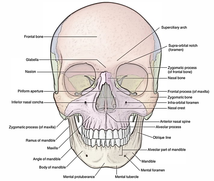
As the name implies, an articulation is where two bone surfaces come together (articulus = “joint”).

There are three general classes of bone markings: (1) articulations, (2) projections, and (3) holes. describes the bone markings, which are illustrated in ( ). The surface features of bones vary considerably, depending on the function and location in the body. In this region, the epiphyses are covered with articular cartilage, a thin layer of cartilage that reduces friction and acts as a shock absorber. The periosteum covers the entire outer surface except where the epiphyses meet other bones to form joints ( ). Tendons and ligaments also attach to bones at the periosteum. The periosteum contains blood vessels, nerves, and lymphatic vessels that nourish compact bone. The outer surface of the bone is covered with a fibrous membrane called the periosteum (peri – = “around” or “surrounding”). The medullary cavity has a delicate membranous lining called the endosteum (end- = “inside” oste- = “bone”), where bone growth, repair, and remodeling occur. When the bone stops growing in early adulthood (approximately 18–21 years), the cartilage is replaced by osseous tissue and the epiphyseal plate becomes an epiphyseal line. Each epiphysis meets the diaphysis at the metaphysis, the narrow area that contains the epiphyseal plate (growth plate), a layer of hyaline (transparent) cartilage in a growing bone.

Red marrow fills the spaces in the spongy bone. The wider section at each end of the bone is called the epiphysis (plural = epiphyses), which is filled with spongy bone. The skull reaches its definitive size around the 20 th year of life.A typical long bone shows the gross anatomical characteristics of bone. Most features of the adult skull appear during the first two years of life, including the inner and outer tables, diploic space, vascular markings, and grooves for the dural sinuses. During birth, the sutures and fontanels overlap (molding) to aid the head's passage through the birth canal. The posterior fontanel closes before the age of 3 months and may already be closed at birth, while the anterior fontanel closes after the age of 18 months this is of prime importance for performing cranial ultrasound. two posterolateral (or mastoid) fontanelles.two anterolateral (sphenoidal) fontanelles.In addition to the sutures, there are six fontanelles (also spelled fontanels) which are six flat membranous junctions where three or four sutures meet: The sagittal suture develops from neural crest cells and the coronal suture, from paraxial mesoderm. The plates are separated by broad dense connective tissue seams, the sutures. The calvarial bones form within a collagen matrix as bone spicules, which radiate from a primary ossification center in each bony plate toward the periphery.

The mesoderm undergoes direct intramembranous ossification from which the flat calvarial bones are formed, that is to say, there is no intervening cartilaginous scaffolding. The membranous neurocranium develops from paraxial mesoderm and neural crest cells. The cranial vault develops from the membranous neurocranium. the interparietal part of the occipital bone.the squamous part of the paired temporal bones.The cranial vault consists of the following flat bones:


 0 kommentar(er)
